Medipix - High Energy Physics collaborators deliver technological breakthrough behind world’s most advanced X-ray detector
Submitting Institution
University of GlasgowUnit of Assessment
PhysicsSummary Impact Type
TechnologicalResearch Subject Area(s)
Physical Sciences: Other Physical Sciences
Information and Computing Sciences: Artificial Intelligence and Image Processing
Summary of the impact
Medipix-based detectors are the best pixelated X-ray detectors available
on the market and are commercialised by PANalytical under the brand name
PIXcel. At the core of PIXcel is the Medipix2 chip, which was developed
around a photon counting breakthrough conceived by the Medipix
collaboration and is unique in its adaptability, high spatial resolution,
high dynamic range and low noise. This product is the direct result of an
exclusive license and a collaboration agreement between PANalytical and
the Medipix collaboration, coordinated by CERN and comprising a further
sixteen leading physics research institutes in Europe. The University of
Glasgow is the only UK institution to be one of the four founding members
of the Medipix1 collaboration.
Underpinning research
In the 1990's, a University of Glasgow team led by Prof Kenway Smith FRSE
was actively involved in the development of hybrid pixel detectors for the
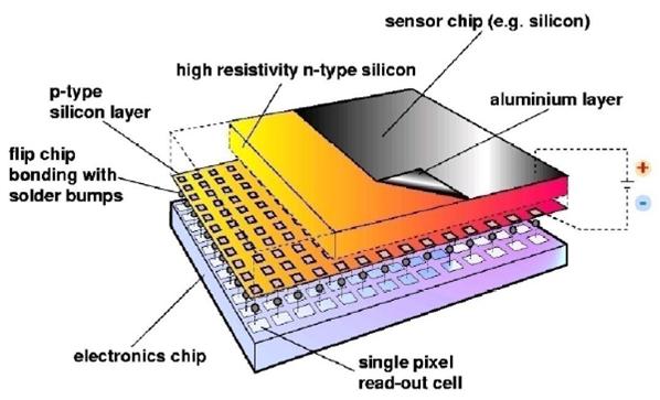
Large Hadron Collider (LHC). These hybrid pixel detectors combine a CMOS
chip and an array of readout electronics channels, connected via micro
bump bonds to an identically segmented sensor chip. The primary motivation
was to develop a 2-D detector capable of time stamping high energy physics
(HEP) events at the expected 40MHz collision rate of the LHC. In the
course of this research, it became clear that such a technology could also
be useful in medical imaging [1].
The Glasgow group identified the need for new technology in X-ray imaging
as being particularly significant. In photon counting mode, the Medipix
chip could deliver significant advances in the detection of X-ray
diffraction patterns. The Glasgow team demonstrated this for the first
time, using hybrid pixel detectors at a synchrotron X-ray source [2]. The
Omega3 chip, a predecessor of Medipix, was used in this early work, and it
was repeated with the Medipix chip when it became available.
However, to be useful for medical and other imaging applications, the
electronics readout chip would have to be adapted to produce X-ray images
which are high resolution and noise-free. From the starting point of this
pioneering Medipix1 chip in 1997 grew the design brief of what was to
become the Medipix2 chip produced in 2001 and now at the core of the
PIXcel detector. This brief was outlined by the Glasgow research team
including Dr R Bates (Research Associate 1999-2007 and Research Fellow
2007-present), Dr S D'Auria (Research Associate 1999-2010 and Research
Fellow 2010-present) and Dr C Raine (Research Assistance 1980-98), along
with teams from Freiburg, Pisa and the European Organization for Nuclear
Research (CERN). The Glasgow group effort was led by Prof K Smith
(Lecturer/Reader 1963-93, Professor 1993-2003) from 1991 until 2003 and
since then by Prof V O'Shea (Research Associate 1992-2005, Lecturer
2005-2010, Professor 2010-present). Glasgow brought design engineering
expertise to the Medipix ASIC design group and performed much of the
initial characterisation of the Medipix1 chip.
As interest in medical imaging applications grew and the collaboration
expanded, it became possible to consider a more advanced chip design
involving simultaneously shrinking the pixel size, increasing its
complexity and increasing the number of pixels, which resulted in the
Medipix2 chip. Seventeen institutions contributed ideas to the Medipix2
readout chips, pixel sensors design, characterisation of the pixel modules
and applications of the Medipix2 chips. The Glasgow team contributed to
the chip design, including the features necessary to make it an efficient,
high-resolution photon-counting device. The Glasgow team also tested and
characterised the Medipix2 chip, studying novel sensor materials (GaAs,
CdTe) and structures (3-D Si) for a variety of applications. As a
consequence of these and other studies [3], further developments of the
technology were made resulting in the Medipix3 chip. Today, the Medipix
family of hybrid pixel detectors are used for medical imaging including
computer tomography scans, material analysis including pharmaceuticals,
nuclear power plant decommissioning and astronomical adaptive optics.

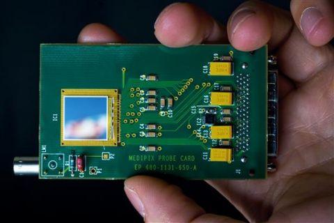
 Figure 1: A picture and schematic diagram of the high-sensitivity, low-noise Medipix2 chip.
References to the research
[1] C Da Via, et al, "Gallium arsenide pixel detectors for medical
imaging", * Nuclear Instruments and Methods A, Volume 395 (1997) 148-151.
DOI:
10.1016/S0168-9002(97)00631-1
Glasgow Authors: C Da Via, R Bates, C Raine, V O'Shea, KM Smith
Description: This paper is one of the first papers to use a hybrid
pixel detector assembly for non- HEP applications using the fore-runner
LHC1/Omega3 chip. It demonstrates that such devices can be used for a
variety of applications, from medical imaging to DNA sequencing.
Glasgow contribution: These were in the specification of the chip
and the design, evaluation and analysis of the device characterisation.
Chip characterisation is a vital part of the design phase, proving that
the device works as expected and feeding back to the design.
[2] S Manolopoulos, et al, "X-ray powder diffraction with hybrid
semiconductor pixel detectors", * Journal of Synchrotron Radiation, Volume
6 (1999), 112-115.
DOI:
10.1107/S0909049599001107
Glasgow Authors: S Manolopoulos, R Bates, V O'Shea, C Raine, KM Smith
Description: This paper demonstrates the first use of a hybrid pixel
detector in a Synchrotron beam line. The detector is the Omega3 chip. The
experiment was repeated with the Medipix1 chip when this became available.
Glasgow Contribution: The Glasgow group defined the experiment,
performed the experiment with help from the beam line scientists, and
performed the data analysis.
Glasgow authors: K Mathieson, MS Passmore, V O'Shea, RL Bates, KM
Smith, M Rahman Description: This paper models charge sharing in
hybrid pixel detectors. The models are developed and benchmarked against
an analogue pixel sensor with large pixel size. The consequences of charge
sharing are then developed for the smaller 55µm x 55µm sized pixels of the
Medipix2 chip. The results of this paper directed the attention of the
Medipix collaboration towards the need for the Medipix3 architecture with
attempts to mitigate charge sharing effects.
Glasgow contribution: The simulations and experimental work was
all performed in Glasgow.
The non-Glasgow authors developed and produced the analogue pixel
detector.
Details of the impact
Physicists at Glasgow first recognised the potential medical applications
of the hybrid particle detector in the 1990's. In the years that followed,
the Medipix collaboration developed an innovative new microchip, and then
brought the technology to market through a commercialisation deal with
leading X-ray equipment manufacturer PANalytical. The resulting PIXcel
range is the most advanced technology of its kind on the market, offering
a cost-effective, accessible, but highly adaptable machine producing
images of unrivalled quality.
The Head of the Knowledge Transfer Group at CERN said: "At CERN we assess
the impact of knowledge transfer projects in terms of the number of people
benefitting, project visibility, and wider dissemination as well as
financial return. In these terms Medipix2 and its relationship with
Panalytical is one of the projects with highest impact that CERN has been
involved with."
PANalytical, formerly known as Philips Analytical, is one of the leading
suppliers of X-ray materials analysis equipment. The company offers a
range of X-Ray Diffraction (XRD) instruments, which are sold to customers
worldwide both in academia and industry. XRD is a non-destructive
analytical technique used to determine the crystalline structure and
elemental composition of materials. Its applications range from
pharmaceuticals and petrochemical to semiconductor, electronics, and
nanotechnology. XRD is a particularly versatile technique as it can
provide information on the chemical and physical characteristics of a
range of materials including powders, thin films or bulk materials.
The Medipix2 collaboration secured a Collaboration Agreement with
PANalytical in 2001, and the company supported the development of the
Medipix2 chip in return for exclusive rights to commercialise the
resulting technology in its new Empyrean product line. The company
successfully launched the first PIXcel product in 2007. Today, PANalytical
sells three different products based on the Medipix2 pixelated detector.
The introduction of a series of PIXcel detectors (PIXcel1D,
PIXcel3D and PIXcel3D
2x2) has added new dimensions to XRD analysis. With this complete series
of detectors users can obtain the best available data quality. This
technology breakthrough required sustained input for approximately 8 years
from the Medipix collaboration before yielding mature, marketable
products.
PANalytical's PIXcel, offers a number of distinct advantages over its
competitors. First, it is uniquely adaptable, being the only detector on
the market that can operate as a point-, linear-, 2-D or 3-D detector, all
in one. This adaptability is complemented by its high spatial resolution,
high dynamic range and low noise, which make PIXcel the most advanced
photon counting detector in the world.
The PIXcel3D X-ray detector offers flexibility
and performance that sets it apart from all competing systems, which use
separate detectors to cover the same range, thus increasing the cost.
PIXcel's all-in-one capabilities are more cost-effective, allowing modes
that were so far only available at large synchrotron facilities. In 2011,
Empyrean, the product line of XRD systems incorporating PIXcel, won an
award for the best technology innovation from R&D Magazine. It
continues to be a very successful product line that is well received by
customers.
In 2013, PANalytical introduced a further innovation to its Empyrean
product line: the ability to perform grazing incidence small-angle X-ray
scattering (GISAXS). GISAXS is a technique that is used to investigate the
physical and chemical properties of nanostructured thin films. However, as
an advanced analytical technique, it is traditionally restricted to users
of synchrotron beam lines.
Thanks to the high efficiency and low noise of the Medipix2 detector,
high quality images can be obtained even using laboratory sources with
intensity orders of magnitude lower than that from a synchrotron beam. As
nanomaterials become more widespread in a number of applications,
affordable and convenient access to this technology is of great advantage
to users both in academia and industry.
The PANalytical XRD Product Marketing Manager said: "We are very proud to
be a commercial partner in Medipix2. PANalytical has a track record of
innovation and this association will keep us at the forefront of detector
technology for many years to come. We are determined to continue
developing the best, and most innovative, detectors for all our future XRD
customers. Empyrean is PANalytical's answer to the challenges of modern
materials research. While today's research themes are nanomaterials, life
sciences and renewable energy, tomorrow's science may move in a totally
different direction. The lifetime of a PANalytical diffractometer
stretches further than the typical horizon of a single research program
and for many scientists, an ability to accommodate change is a 'must-have'
feature in their decision to invest in an XRD system."
Sources to corroborate the impact
Medipix Knowledge Transfer
http://knowledgetransfer.web.cern.ch/life-sciences/from-physics-to-medicine/medipix
PANalytical Website Pixcel description
http://www.panalytical.com/Empyrean/Features/PIXcel.htm
PANalytial Empyrean brochure
Multi-Purpose X-Ray Diffractometer (XRD) and Computer Tomography (CT) with
the PANalytical Empyrean
http://www.azom.com/materials-video-details.aspx?VidID=657
X-ray measures in multiple dimensions
http://www.rdmag.com/Awards/Rd-100-Awards/2011/08/X-ray-measures-in-multiple-dimensions/
Innovations in Pharmaceutical Technology
http://www.iptonline.com/newproduct.asp?id=1123
PANalytical — New GISAXS option on Empyrean
http://www.panalytical.com/News/New-GISAXS-option-on-Empyrean.htm
Newswire & PR — PANalytical Demonstrates New GISAXS Option on
Empyrean
http://www.prnewswire.com/news-releases/panalyticals-empyrean-to-be-featured-at-intersolar-north-america---the-world-of-xrd-analysis-is-no-longer-flat-97929394.html
Head of the Knowledge Transfer Group, CERN, European Organization for
Nuclear Research, can provide corroboration of the impact of the
commercial relationship with PANalytical for CERN and the broader
importance of the impact of the Medipix project.