UOA15-05: Imaging software for cancer diagnosis
Submitting Institution
University of OxfordUnit of Assessment
General EngineeringSummary Impact Type
TechnologicalResearch Subject Area(s)
Physical Sciences: Other Physical Sciences
Information and Computing Sciences: Artificial Intelligence and Image Processing
Medical and Health Sciences: Neurosciences
Summary of the impact
Key advances in the earlier diagnosis of cancer, leading to better
treatment and higher survival rates, have resulted from the
commercialisation of unique imaging software that exploits research from
the Department of Engineering Science. The software products that came
from this research, Volpara™, XD and XRT are now used at major cancer
centres worldwide (with approximately 1100 software installations), aiding
treatment of tens of thousands of patients every year. Between 2009 and
July 2013, Volpara™ scanned over 1.2 million mammograms, enabling the
early detection of around 1800 cancers. The products' success has
catalysed significant improvements in cancer care, and generated an
estimated £9M in sales over the past two years for the spinout companies
established to develop them (Matakina, based in New Zealand, and Mirada
Medical, based in the UK).
Underpinning research
Whilst the key concepts behind these life-saving products were at first
controversial, they are now firmly established as underpinning
quantitative mammographic techniques. The beginnings of the work go back
to 1989, and a key phase was Ralph Highnam's postdoctoral work 1992-1999
[1, 2, 3], with further developments continuing since then. The research
was led by Prof. Mike Brady, (Professor of Information Engineering,
1985-2010 and Professor of Oncological Imaging 2012-present). The
research underpinning the advances achieved is summarised below:
-
Mammography imaging and analysis. A key problem, prior to
Brady's work, was that mammography systems relied on human perception
and judgement. The image in a mammogram is affected by X-ray machine
parameters as well as the breast tissue through which the X-rays pass.
But conventionally, information about parameters such as exposure time,
tube voltage (set by the user) and anode type (which varies from machine
to machine) was recorded inaccurately, for digital mammograms, or even
not at all, for film-screen mammograms. The result was that tissue from
the same breast might appear to have changed in images taken a short
time apart — making accurate analysis of the tissue much more difficult.
This subjectivity led to inconsistent clinical assessments of patients.
Working with Dr Ralph Highnam (PDRA 1992-1999 and later co-founder and
CEO of Matakina), Brady incorporated explicit mathematical models of the
physics of image acquisition into medical image analysis, as well as
taking account of the 3D nature of this problem. This approach underpins
Matakina's Volpara™ product, which estimates breast tissue density from
conventional mammography scans and produces a simple-to-read `score'
giving an accurate indication of this key breast cancer risk factor [1, 2,
3, 4]. As a mammogram is a 2D image of the 3D breast, so information in
the third dimension (i.e. perpendicular to the compression plates)
is inevitably lost. Brady and Highnam discovered that, starting from
estimates of various, often unknown parameters, their model's predictions
could be matched against the mammogram and adjusted until a best fit was
achieved. This provided a way of estimating, at the pixel level, the
amount of dense tissue in the apparently lost third dimension and
measuring the amount of non-fat tissue in the breast — enabling images of
tumours to be enhanced.
This model-based approach, which makes it much easier to analyse the
amount of dense tissue as it varies from pixel to pixel, has transformed
mammogram analysis, enabling those at greatest risk of developing breast
cancer to be identified much more easily and, in many cases, much earlier
than previously possible (see Section 4). While Matakina and other
companies such as GE Healthcare and Hologic have developed software
systems based on this research, Brady and Highnam have continued to refine
their mathematical model to extend its capabilities and further refine its
clinical value [5, 6].
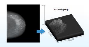
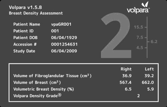
Volpara™ calculates a patient's breast tissue
density automatically and objectively from digital mammography data
(left), and then presents it the results in an easy-to-read format
(right)
-
Mirada Solutions' technology: XD and XRT are firmly rooted in
the scientific insights and implementation methods developed by Brady
and his colleagues. The focus is on the concept of `image registration'
which concerns the alignment of images, typically of different types
acquired at different times and different spatial resolutions.
The challenge of image registration was made significantly more complex
by the emergence, around the year 2000, of combined PET-CT machines — and
specifically by the drive to fuse separate 3D PET (positron emission
tomography) and CT (computed tomography) images into a single, better
quality image. Aligning a PET image of a patient's entire body with a CT
image of a chest cavity or other body part required a painstaking
computing process typically taking 1-2 hours to complete. Building on
work Brady conducted in the 1990s on deformable registration of images of
different types, Mirada Solutions successfully developed an image
alignment method taking just 5 minutes, and seldom requiring the user's
intervention. Since this breakthrough, the company has continued to
harness and apply a stream of scientific advances achieved by Brady and
his team (e.g. in dosimetry for X-radiation therapy).
References to the research
(best indicators of research quality are marked `Q')
1. Brady, J.M. and Highnam, R.P. `Mammographic Image Analysis' (1999).
Kluwer Series on Medical Image Understanding. ISBN 0-7923-5620-9. `Q'
Monograph outlining the mammography methods pioneered by Brady and
Highnam, as well as their work on MRI and PET analysis of breast images.
The two above papers set out Brady and Highnam's model of mammographic
image formation.
4. Behrenbruch, C.P., Marias, K., Armitage, P.A., Yam, M., Moore, N.,
English, R.E., Clarke, P.J. and Brady, J.M. `Fusion of Contrast-Enhanced
Breast MR and Mammographic Imaging Data' (2003). Medical Image
Analysis, 7(3), pp 311-340, Elsevier.
http://dx.doi.org/10.1016/S1361-8415(03)00015-X
Although much of Mirada Solutions' work on image registration was
confidential, this paper summarised the methods and showed a number of
fusion results in the case of breast imaging.
5. Highnam, R.P., Pan, X., Warren, R., Jeffreys, M., Davey Smith, G. and
Brady, J.M., `Breast Composition Measurements Using Retrospective Standard
Mammogram Form (SMF)' (2006). Physics in Medicine and Biology, 51,
pp 2695-2713
http://dx.doi.org/10.1088/0031-9155/51/11/001
`Q'
Extend Brady and Highnam's method and makes it applicable to all
mammograms.
6. Highnam, R.P, Brady, J.M, Yaffe, M.J., Karssemeijer, N. and Harvey, J.
`Robust Breast Composition Measurement — Volpara™' (2010). Digital
Mammography, Springer Lecture Notes in Computer Science, 6136, pp
342-349.
http://link.springer.com/chapter/10.1007%2F978-3-642-13666-5_46
The methods outlined in Refs. 1-5 address the general physics of
mammography; Ref. 6 focusses on issues more specific to the Volpara™
product.
Grants in support of this research:
• Cancer Research UK: Quantitative Assessment of Mammograms, 2001-2004,
£113,980. Principal Investigator: Mike Brady.
• EPSRC: From Medical Images and Signals to Clinical Information,
2001-2007, £1,986,920 (Oxford component) (ref: GR/N14248/01). Principal
Investigator: Mike Brady.
• EPSRC: eDiamond Grid Project, 2002-2005, £1,290,367 (award to
investigate breast cancer) (ref: GR/S20956/01). Principal Investigator:
Mike Brady.
• EPSRC: Investigating a Model-based Approach to Breast Imaging,
2008-2010, £389,273 (ref: EP/E031978/1). Principal Investigator: Mike
Brady.
• EU FP7: ASSURE (Adapting Breast Cancer Screening Strategy Using
Personalised Risk Estimation), 2012-2015, €5.6M. Principal Investigator:
Nico Karssemeijer (University of Nijmegen).
Details of the impact
Brady's research has triggered the development of products that are
having a direct impact on cancer prevention, detection and treatment
regimes. By saving lives and simplifying clinical practice, the benefits
accrue to millions of patients and thousands of healthcare professionals
worldwide. Working with colleagues in academia and industry, Brady set up
the two spin-out companies to exploit the potential of the research, and
he continues to work closely with them:
-
Mirada Solutions: formed in 2000 from a merger of two previous
spinouts established by Brady (OMIA and OXIVA), this firm focused on
image fusion and underwent a series of acquisitions and name-changes
before Brady and three former colleagues bought it back. This led to the
formation of Mirada Medical (http://www.mirada-medical.com)
in 2009, which is based in Oxford and specialises in cancer diagnosis
and radiation planning software.
-
Matakina (http://www.matakina.com):
set up in 2009 to exploit Brady and Highnam's breakthroughs in
mammography software, this company also harnesses the outstanding
research skills of Professor Nico Karssemeijer (University of Nijmegen,
the Netherlands) and Professor Martin Yaffe (University of Toronto,
Canada).
Health Impact — earlier diagnosis of cancer
Women with very dense breast tissue have a 4-6 times higher risk of
developing breast cancer than those with tissue consisting predominantly
of low-density fat. Uniquely, Volpara™ converts any digital breast
mammogram into a `normalised' image, presented to a clinician as a summary
screen (see photo on p. 2). Thanks to these capabilities, the software is
used as a time-saving device in cancer screening worldwide. In summary,
Volpara™ has substantially changed clinical practice in the hospitals
using it. Endorsements from clinical practitioners include:
- "Volpara was implemented here since October 2010, and we have found it
to be accurate and reliable" [7].
- "Volpara is easy to use with minimal training" [8].
- "Volpara has made the whole [mammogram] process much more streamlined
and convenient for women" [9].
- "Volpara has been used in a large, screening trial because of its
robust clinical record" [10].
Mirada Medical's software is delivering quicker, easier, more effective
diagnosis and monitoring for a range of additional cancers, including lung
cancer, melanoma, liver cancer and head and neck cancer.
Economic Impact — wealth creation and job generation
As of July 2013, Matakina's Volpara™ software is being used in Australia,
Belgium, Canada, Chile, Denmark, Finland, Ireland, Italy, Japan, Malaysia,
the Netherlands, New Zealand, Norway, Saudi Arabia, South Africa, South
Korea, Sweden, Switzerland, Thailand and the US, as well as the UK. In
2012 alone, over 500,000 women had their breast density assessed using the
software — including 72,000 at Eastern Radiology in Greenville, North
Carolina, 50,000 at Radboud Hospital in Nijmegen and 48,000 at the Samsung
Medical Center in Seoul, South Korea [11].
Mirada Medical's software is used at all major cancer centres in the US
(e.g. the Johns Hopkins University Hospital in Baltimore, Maryland, and
the MD Anderson Cancer Center in Houston, Texas), in the UK (e.g. the
Oxford University Hospitals NHS Trust and the Western General Hospital in
Edinburgh) and in Denmark, India and the Netherlands. In addition, the
firm sells its products to companies such as Toshiba, Siemens, Vital,
Carestream, McKesson and Sectra who incorporate them into their own
products. Major equipment suppliers Varian and GE Healthcare have also
placed contracts with the company to develop innovative software.
This extensive worldwide take-up of the spinouts' range of software
products has translated directly into impressive commercial performance.
Matakina has 116 installations in 20 countries and Mirada has around 1000
installations in 10 countries, with a combined annual turnover now around
£5 million. Such figures highlight the fact that Brady's pioneering
research has ultimately had a significant global impact not only in
healthcare but also in economic terms.
Sources to corroborate the impact
7. M.D., Eastern Radiology, North Carolina. Corroborates Volpara™ being
accurate and reliable. Testimonial at http://www.volparasolutions.com
8. M.D., Jules Bordet Institute, Belgium. Corroborates Volpara™
is easy to use with minimal training. Testimonial at http://www.volparasolutions.com
9. Radiologist, Sutter Health, US. Corroborates the impact of Volpara™
being more convenient. Testimonial at http://www.volparasolutions.com
10. Associate Professor of Clinical Epidemiology, University Medical
Centre, Utrecht, the Netherlands. Corroborates the utility of Volpara™.
Testimonial at http://www.volparasolutions.com
11. CEO, Matakina. Corroborates widespread uptake Volpara™
internationally.