Optimising Gradient and Shim Coils for Next-Generation Magnetic Resonance Imaging Systems
Submitting Institution
University of NottinghamUnit of Assessment
PhysicsSummary Impact Type
TechnologicalResearch Subject Area(s)
Physical Sciences: Other Physical Sciences
Engineering: Biomedical Engineering
Medical and Health Sciences: Neurosciences
Summary of the impact
Theoretical and computational methods for optimising the design of
gradient and shim coils with
arbitrary shapes and topologies were developed in collaboration with
Magnex Scientific as part of a
CASE award (2004-07). The resulting software was licenced to Agilent (who
now own Magnex
Scientific), for whom it has opened up new market opportunities in the
supply of novel magnetic
resonance imaging systems, leading to £3.4M sales since 2009. The software
has also been used
by Paramed Medical Systems to improve their `open' magnetic resonance
imaging systems, which
are optimised for orthopaedic imaging, allow vertical subject posture, and
facilitate image-guided
treatment, as well as offering a better patient experience. Our work has
thus resulted in impact in
the economy and healthcare.
Underpinning research
The use of gradient and shim coils is integral to the operation of
magnetic resonance imaging
(MRI) systems. The three gradient coils found in each scanner are used to
produce magnetic fields
that vary linearly with position along three orthogonal axes. These field
gradients are crucial for the
spatial encoding of the magnetic resonance signal — a process which forms
the basis of MRI.
Gradient coil performance strongly influences many aspects of magnetic
resonance (MR) image
quality, including the spatial resolution and achievable contrast. Shim
coils are used to correct
unavoidable inhomogeneities in the main static magnetic field that is also
used in MRI. These
inhomogeneities may arise from various sources, such as the magnetic
susceptibility of a human
subject, or imperfections in the magnet construction, and, if uncorrected,
result in significant image
distortions.
The Nottingham MRI group has a longstanding track record in the
development of improved
hardware for MRI systems, dating back to the early work for which Sir
Peter Mansfield shared the
2003 Nobel Prize in Medicine or Physiology. This research activity has
continued to the present
day, with a particular emphasis on gradient and shim coil technology.
Previous high impact work in
this area includes the development in the late 1980s of actively-shielded
gradient coils, which are
now used in all MR scanners, and the subsequent establishment of methods
for designing high
efficiency coils. These developments formed the basis for the substantial
improvement in gradient
system performance in the 1990s, which first allowed the routine
implementation of functional
magnetic resonance imaging of the brain and other imaging procedures that
rely on strong, rapidly
switched gradients.
Since the late 1990s, the focus of our research has shifted to the
development of methods for the
design of coils with complex geometries or topologies that are required to
enable new applications
and advances in MRI. Until recently, gradient coils for MR scanners were
designed using a simple
parameterisation of the current density that represents the energised coil
windings. This approach
is well suited to the production of gradient coils that are wound on
simple surfaces, such as the
long cylinders used in early MR scanners, but cannot easily be applied to
designing coils on more
complex surfaces that may be truncated, folded, gapped or split. Research
into these complex coil
shapes was motivated by their requirement in novel MRI systems, including:
- scanners incorporating insert gradient and shim coils that can be used
for enhanced
imaging of particular target structures — for example, insert head
gradient and shim coils
that fit closely around the head
- `open' MRI scanners which depart from the conventional tubular shape —
these can
enhance the patient experience through reduction of stress (particularly
important for
paediatric and obese patients) and can also be used for real-time
monitoring of surgery and
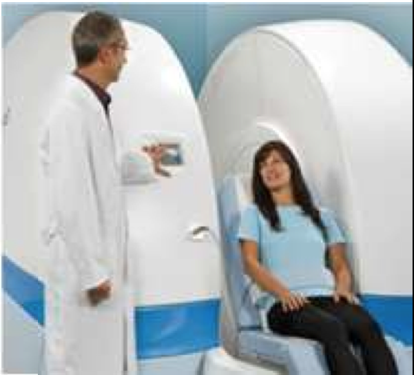
other complex processes; an example is the `MROpen' system, developed by
Paramed
Medical Systems (see Figure), which allows imaging of human subjects in
weight-bearing,
vertical, postures
- hybrid systems, such as combined positron emission tomography (PET)
and MRI scanners,
and combined radiotherapy and MRI systems, in which the coils must be
designed to be
compatible with the spatial arrangement and operation of the equipment
required for the
additional modality.
Boundary element methods (BEM) offer a powerful approach for coil design,
allowing the
generation of coils wound on arbitrarily-shaped surfaces, so as to produce
any form of field
variation that is consistent with Maxwell's equations. This approach
involves meshing the current-carrying
surface into an array of boundary elements and then setting the current
density at each
element, so as to minimise a functional that reflects the desired coil
characteristics.
Between 2004 and 2007, Bowtell (a member of the Nottingham MRI
group), working with CASE
Ph.D. student, Poole, applied the boundary element approach to the inverse
problem of designing
coils on arbitrarily shaped surfaces that would produce specified magnetic
field distributions. The
outcome was a novel inverse BEM (IBEM) approach. This was implemented in
software written by
Poole, and combined with a powerful mesh-generating program, so as to
allow the design and
production of coils with completely arbitrary geometry [1]. Poole's
studentship was funded by
Magnex Scientific/Varian Inc.[i] and his Ph.D. work linked into an
EPSRC-funded project which
focused on applying techniques in computational mechanics to the design
and analysis of gradient
and radio-frequency coils for MRI [ii].
In 2007, Poole and Bowtell worked with Alun Lucas, Rob
Hawkes and Adrian Carpenter (Wolfson Brain Imaging
Centre at the Addenbrooke's Hospital in Cambridge), and
Dan Green and Simon Pittard (Varian Inc., now Agilent), on
the design and construction of coils for a hybrid PET-MRI
system [2]. Combining PET and MRI within one instrument
requires many engineering compromises as the equipment
for the two modalities vies for the space closest to the
sample/subject. Nevertheless, a combined PET-MRI
system offers many potential benefits for clinical and pre-clinical
imaging. In the configuration which was developed,
the PET detectors reside in a gap between the two halves
of a 1T split-magnet cryostat. The gradient and shim coils
had to incorporate a 110 mm gap from which wires are
excluded so as to avoid compromising the process of
positron detection. It was not possible to produce coils with
this gapped geometry using conventional methods of coil
design, but Poole's IBEM software was able to cope with
the complex coil shapes. It was used to design three,
orthogonal, (magnetically) shielded gradient coils and a
shielded, zero-order shim coil. These coils were
constructed and tested in the hybrid PET-MRI system and
successfully used in simultaneous PET-MRI experiments
[2].
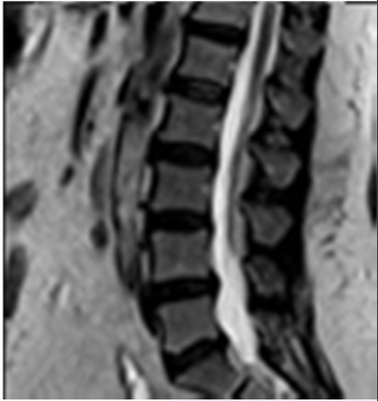
 Top: Lumbar spine MRI scan (imaged under vertical compression)
 Left: Paramed’s MROpen scanner.
References to the research
(*denotes paper which best describes quality of research)
1) *M. Poole and R. Bowtell, `Novel gradient coils designed
using a boundary element method.',
Concepts in Magnetic Resonance Part B 31B, 162 (2007).
DOI: 10.1002/cmr.b.20091
2) *M. Poole, R. Bowtell, D. Green, S. Pittard, A. Lucas, R. Hawkes, A
Carpenter, `Split
gradient coils for simultaneous PET-MRI', Magnetic Resonance in
Medicine 62,1106 (2009).
DOI: 10.1002/mrm.22143
3) *R. Bowtell and R. M. Bowley `Analytic calculations of the
E-fields induced by time-varying
magnetic fields generated by cylindrical gradient coils', Magnetic
Resonance in Medicine 44, 782
(2000).
DOI: 10.1002/1522-2594
4) R. Bowtell and A. Peters `Analytic approach to the design
of transverse gradient coils with co-axial
return paths', Magnetic Resonance in Medicine 41, 600
(1999).
DOI: 10.1002/(SICI)1522-2594
5) P. Mansfield, B. L. W. Chapman, R. Bowtell, P. Glover, R. Coxon,
P. R. Harvey, `Active
acoustic screening — reduction of noise in gradient coils by Lorentz
force balancing', Magnetic
Resonance in Medicine 33, 276 (1995).
DOI: 10.1002/mrm.1910330220
Grants
i. Magnex CASE studentship (01/10/2003-31/03/2007) £22,000
ii. `Forward & inverse analysis of electromagnetic fields
for MRI using computational mechanics
techniques' I.A. Jones, R. Bowtell, A. Becker, P.M. Glover, H.
Power, EPSRC Grant
GR/T22445/01 (1/2/05-3/1/2008) £208,925
Details of the impact
The strong track record of the MRI group in the development of gradient
coil technology for
magnetic resonance scanners led to the sponsorship by Magnex Scientific
(manufacturer of
magnets, gradient coils and shim coils for MRI) of a CASE studentship for
Poole under the
supervision of Bowtell. Poole's Ph.D. project, which commenced in
2004, focused on the
development of insert gradient and shim coils for use in high-field MRI of
the human head. The
IBEM approach was developed as part of this work, so as to allow the
design of coils wound on
surfaces that fit more closely to the head than the body-sized cylinders
used in most MRI systems.
From 2005-2006, Poole developed a versatile suite of algorithms and
subsequently an IBEM
software package for designing gradient and shim coils on
arbitrarily-shaped surfaces.
This software was demonstrated to our collaborators at Magnex Scientific
during regular project
meetings in 2006 (by this time, Magnex had been bought by Varian Inc.;
Varian were then bought
by Agilent in 2010 for $1.5bn; the manufacturing base has remained in
Yarnton, Oxfordshire
throughout the changes in ownership). It rapidly become clear to Varian
that the IBEM software
offered much greater versatility than the coil design programmes that they
were then using. The
facility to design and produce coils on gapped or split surfaces generated
particular interest. To
demonstrate the potential of the IBEM software, Poole worked with Dan
Green (Varian) to
generate novel, gapped, designs of shielded gradient and shim coils for a
prototype hybrid
PET/MRI system. These coils could not be designed using the software that
Varian were using at
that time.
Subsequently, Varian constructed these coils and supplied them
commercially to Adrian Carpenter
and colleagues at the Wolfson Brain Imaging Centre in Addenbrooke's
Hospital (2008), paying a
3% royalty to the University of Nottingham. Bowtell and Poole
published a paper [2] jointly with
Carpenter's group and the Varian collaborators, describing the novel coil
designs and the first
experimental tests of their performance in the hybrid PET/MRI system.
Having seen the value of
the IBEM approach, Varian extended the agreement with the University of
Nottingham in 2007, and
this now allows them to use Poole's advanced design software, provided
that they pay a 3%
royalty on any coil sales arising from its use. Since 2008, Varian, and
subsequently Agilent, have
used the software to design a number of coil systems, including short head
gradient coils for ultra-high
field imaging, and gapped systems for use in combined radiotherapy/MRI
systems. Agilent's
Research and Development Manager for Gradient Coils [A] describes the
impact on their business:
"The ability of the software to account for complex gradient coil
geometries allows us to explore the
more challenging design aspects of bespoke and integrated systems, for
instance hybrid MRI/PET
and MRI applications in radiotherapy. Agilent's entry into these new
markets has resulted in some
£2.8 million in new system sales since 2009. We also offer some unique
solutions to our customers
in the university research sector; often their challenging requirements
can only be met by the
flexibility of the IBEM software. Sales for these upgrades are of the
order £600k since 2009.
Exploitation of these new markets would not have been possible without
Nottingham's research
into complex gradient coil design."
In 2009, Bowtell established a new collaboration with Paramed
Medical Systems (Genova, Italy)
who sell `open' MRI scanners. As described in Section 2, open MRI scanners
offer several
significant advantages over the conventional tubular design including:
better patient experience,
particularly for the paediatric, anxious or larger patient; weight bearing
and postural diagnostics in,
for instance, the upper neck (cervical) and lumbar spine cases (see
figure); the potential for real-time
monitoring of surgery. The Paramed systems are based on permanent (0.15T)
and MgB2
superconducting (0.5T) magnet technology, offering a cheaper alternative
to whole body scanners.
A Paramed scanner (MROpen; see Figure) was recently installed in the `Upright
MRI Centre', a
private healthcare centre opened in Leeds in September 2012.
Scanners of this type use bi-planar gradient coils that are mounted on
the magnet pole pieces and
so are in close proximity to large amounts of highly permeable iron. The
new collaboration, which
also involved researchers at the Istituto Italiano di Tecnologia (IIT) in
Genova, focused on adapting
the IBEM approach to take account of the nearby magnetic material when
designing coils, and was
driven by Paramed's need to incorporate active magnetic shielding of their
coils so as to improve
system performance. Following Paramed's purchase of the IBEM software, it
was modified, in
collaboration with Bowtell and IIT researchers, and has
subsequently been used in a number of
development projects and products at Paramed, including their core product
lines, the MROpen
(see figure) and MRJ3300 (now known as MRInspire) scanners. Paramed have
implemented new
design processes as a result of the IBEM software which affect their
complete product range (that
currently provides a turnover of €8M/year) and will support future
projected growth. These details,
and the associated commercial advantages which result, are highlighted by
the Research and
Development Manager at Paramed [B]:
"The availability of the IBEM software, modified by IIT and Paramed
during and after collaboration
with Nottingham School of Physics and Astronomy (since 2009), helped us
to build a new design
tool for optimizing gradient coil performances in presence of iron,
which is an extremely important
advantage for MRI system design"
In summary, our numerical methods for the optimisation of gradient coils
have been adopted by
manufacturers of MRI equipment to enhance performance, functionality and
patient care, thus
generating impact in the economy and healthcare.
Sources to corroborate the impact
(available on request).
A) Letter from Research and Development Manager, Gradient Coils, Agilent.
B) Letter from Research and Development Manager, Paramed s.r.l.