Malarial Retinopathy has Redefined the Diagnosis of Cerebral Malaria and Improved the Management of Coma in African Children
Submitting Institutions
University of Liverpool,
Liverpool School of Tropical MedicineUnit of Assessment
Clinical MedicineSummary Impact Type
HealthResearch Subject Area(s)
Medical and Health Sciences: Clinical Sciences, Medical Microbiology, Neurosciences
Summary of the impact
Since 1997 University of Liverpool (UoL) investigators have led global
research into malarial retinopathy, the fundus features associated with
severe malaria. The work has propelled this phenomenon from little-known
curiosity to an essential component in the diagnosis of cerebral malaria
(CM) and has altered understanding of how CM causes coma and kills. It has
changed medical practice of those diagnosing one of the commonest fatal
diseases in tropical countries. Malarial retinopathy is now considered an
essential clinical feature of CM aiding the appropriate management of coma
in infants. This change in practice has expanded from African research
settings to clinical practice required by WHO guidelines and disseminated
in major clinical textbooks from 2008.
Underpinning research
Malaria kills an estimated 655,000 people a year, 86% of whom are
children under five in Africa. Most die of CM, a complication
characterised by sudden and profound coma, convulsions and death in
10-20%. It is not understood how malaria causes coma and death, but
adherence of infected red cells to the microvascular lining
(sequestration) is thought to be critical [4]. However, malaria is a
common infection and a child may present in coma due to another cause and
be misdiagnosed with CM because of a coincidental malaria infection. This
is surprisingly common occurring in 25-50% of cases.
The key problems that this research addresses are
- Can malarial retinopathy detection improve the diagnosis of severe
malaria?
- Can malarial retinopathy improve understanding of disease processes in
severe malaria which in turn improve care and treatment?
The research to address these questions was conducted by UOL researchers
starting in 1997 at the Malawi field site and Liverpool in collaboration
with the Liverpool School of Tropical Medicine (LSTM),
Malawi-Liverpool-Wellcome Clinical Research Unit (MLW), Malawi College of
Medicine and Blantyre Malaria Project (BMP), Michigan State University,
but the ophthalmic input is almost entirely from UOL. No other research
group has had significant output to address these questions, so the impact
claimed is wholly from UoL research. The principal investigator for this
research is Beare (1999-present, now Hon Snr Lecturer) with Harding
(2003-present, Professor of Clinical Ophthalmology) and Molyneux (Prof
until 2009, now Emeritus Prof).
The research has shown that the detection of malarial retinopathy is the
only reliable diagnostic sign or test for CM [1]. About 25% of Malawian
children who appeared to die of CM had another cause of death at autopsy,
whilst co-infected with malaria. The presence of malarial retinopathy was
the only clinical or laboratory feature which was able to distinguish
malarial coma from other causes. Molyneux initiated, co-directed and
funded this ten year autopsy study, and Beare's analysis, supervised by
Harding, established the specificity (100%) and sensitivity (95%) of
malarial retinopathy.
Studies led by Beare and supervised by Harding and Molyneux, conducted in
collaboration with LSTM, MLW and BMP found that (i) the severity malarial
of retinopathy predicts risk of death better than any other clinical or
laboratory variable in CM [2] and (ii) that patients fulfilling the
traditional case definition of CM without malarial retinopathy have lower
mortality, shorter coma, and are more likely to have a pre-existing
predisposition to epilepsy [2,5]. These insights allow greater prognostic,
as well as diagnostic, accuracy enabling clinicians to focus resources.
In 2006 Beare et al commenced a study of retinal perfusion in CM
using a fundus camera with capacity for retinal angiography. The retina is
central nervous tissue with a similar embryology and structure to the
brain, and with comparable malaria parasite sequestration. The study found
that there are retinal perfusion abnormalities in the majority of patients
with CM [3]. The commonest are multiple zones of ischaemia from
microvascular occlusion. There is also breakdown of the blood-retina
barrier in association with ischaemia; and in a much more profound way
prior to death. This study demonstrated CNS perfusion abnormalities in
vivo in CM for the first time, and altered theories of CM
pathogenesis as it is likely that these abnormalities are also mirrored in
the brain [6]. Understanding of CM pathogenesis has turned towards
ischaemia and blood-tissue breakdown; and away from the previous
hypothesis of the effect of a cytokine and neurotransmitter `storm'. Now
substantiated by MRI studies in children with CM, the next generation of
clinical trials are assessing supportive therapies in cerebral malaria, in
particular therapies to reduce intra-cranial pressure and hypoxic injury.
References to the research
1. Beare NAV, Taylor TE, Harding SP, Lewallen S, Molyneux
ME. Malarial retinopathy: a newly established diagnostic sign in
severe malaria. Am J Trop Med Hyg. 2006;75(5):790- 797. http://www.ajtmh.org/content/75/5/790.long
Citations: 103 Impact factor: 2.59
3. Beare NAV, Harding SP, Taylor TE, Lewallen S, Molyneux
M. Perfusion abnormalities in children with cerebral malaria and
malarial retinopathy. J Inf Dis. 2009;199:263-271. doi:10.1086/595735
Citations: 49 Impact factor: 5.848
4. White VA, Lewallen S, Beare NAV, Molyneux ME, Taylor
TE. Retinal Pathology of Pediatric Cerebral Malaria in Malawi. 2009. PLoS
ONE 4(1): e4317. doi:10.1371/journal.pone.0004317 Citations: 38 Impact
factor: 3.730
5. Lewallen S, Beare NAV, Bronzan R, Molyneux M, Taylor
T. Using malarial retinopathy to classify children with clinically defined
cerebral malaria. Trans Roy Soc Trop Med Hyg. 2008;102(11): 1089-1094 doi:10.1016/j.trstmh.2008.06.014
Citations: Impact factor: 2.16
6. Maude RJ, Dondorp AM, Sayeed AA, Day NPJ, White NJ, Beare NAV.
The Eye in Cerebral Malaria: What Can it Teach Us? Trans Roy Soc Trop Med
Hyg. 2009; 103(7):661-664. doi:10.1016/j.trstmh.2008.11.003
Citations: 14 Impact factor: 2.16 .
Key grants
2005 - 2008. The Wellcome Trust (Ref 075125/Z/04/Z). The eye in
life-threatening malaria: a clinical and clinicopathological study,
£93,239, ME Molyneux, NAV Beare, SP Harding.
2011 - 2014. Wellcome Trust programme grant. Retinal
microvasculature in cerebral malaria, £600k, SP Harding, RS
Heyderman, AG Craig, PS Hiscott, ME Molyneux, TE Taylor, S
Kampondeni, NAV Beare, P Knox, Y Zheng.
Details of the impact
Very few research programmes take a clinical sign from obscurity to the
standard textbook description of a disease. This is exactly what research
by the Department of Eye and Vision Science has done with malarial
retinopathy, and it is all the more remarkable because malaria is a common
and frequently fatal disease.
There are approximately 10 million episodes of cerebral malaria in Africa
a year and examination for malarial retinopathy has the potential to
improve the care of all of these cases. It is plausible that half these
cases, since 2008, have some sort of funduscopy as a consequence of this
UoL research, improving the assessment of prognosis in 5 million and
uncovering misdiagnosis in 1.25million. Patients benefit from improved
diagnosis and better directed treatment. This research has "revealed the
importance of the ocular funduscopic examination in distinguishing "true"
cerebral malaria from "faux" cerebral malaria" [15].
Malarial retinopathy is now included in the description of malaria in
standard medical textbooks which is a key indicator of the impact of this
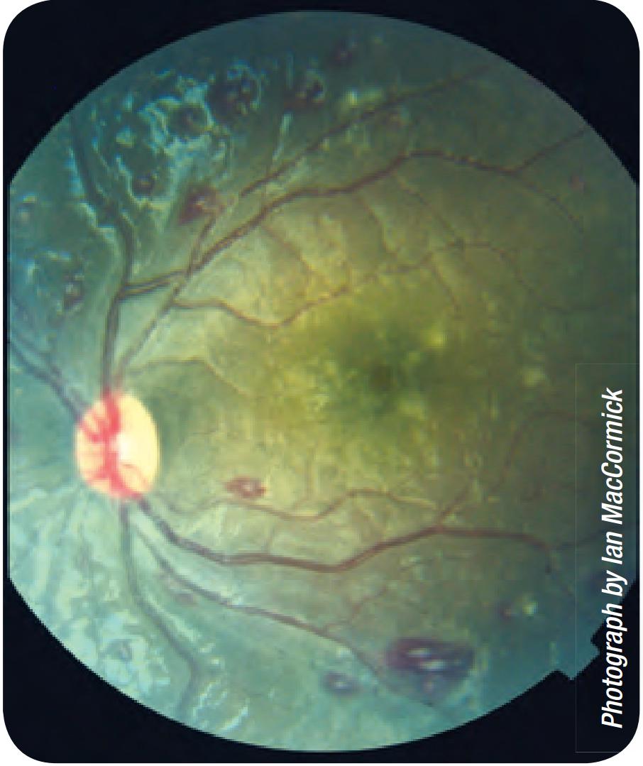
research. Since 2008, retinal photographs taken by Beare and colleagues
such as that presented here) appear in four standard text books -
Harrison's Principles of Internal Medicine [7], Davidson's Principles and
Practice of Medicine [8], Lecture Notes: Tropical Medicine [9] and
Manson's Tropical Diseases[10], as well as multiple reviews on severe
malaria and its pathogenesis. These are major reference works used by
clinician's worldwide to guide clinical practice. Malarial retinopathy
(with the UoL figure) and the importance of funduscopy is now included in
the 2013 WHO guidelines on severe malaria [11], particularly used by
clinicians and policy makers in malaria-endemic countries to determine
best practice, and also regional guidelines eg South-East Asian Regional
Guidelines for the Management of Severe Malaria in Large Hospitals 2006
[12].
 Note the characteristic patchy retinal whitening around the fovea (~3
disc diameters to the right of the optic disc) and also some white-centred
haemorrhages.
The reasons that the authors of these authoritative texts have included
malarial retinopathy in the description of malaria for the first time are
the quality of the UOL research and the importance of its findings. This
includes the startling retinal photographs and results of retinal
angiography which demonstrated graphically the information on CM
pathogenesis that can be gleaned from the retina. The high quality colour
images allowed the features of malarial retinopathy to be demonstrated to
non-ophthalmologists, and to teach them to recognise malarial retinopathy
for themselves. The research has been disseminated by peer-review
publication and conference presentation to key opinion leaders initially
before wider dissemination in textbooks and guidelines. Publications 1 and
2 were featured by the American Academy of Ophthalmology in its EyeNet
magazine and on its Homepage respectively, as well as reported by medical
media and Voice of America radio.
The discovery that malarial retinopathy alone can reliably distinguish
malarial coma from co-infection in a comatose child was of particular
importance, and has literally redefined the disease of cerebral malaria.
Now malarial retinopathy is required to be present in order to diagnose CM
with specificity. This is established practice in Malawi and has been
taken up by other research units in endemic areas; as well as clinical
practice in endemic and non-endemic areas to the benefit of critically-ill
children in coma. This is now confirmed in a popular website, www.malariasite.com
[13].
The Department of Eye and Vision Science has worked with its
collaborators who have set up the Paediatric Research Ward in Malawi and
have provided for patients with severe malaria and other comas from 1990
to present. Since 2008 this has cared for more than 1,500 patients who
have had funduscopy. The malarial retinopathy programme is led by the
Department of Eye and Vision Science, whilst collaborators provide
expertise in malaria.
Improved specificity in the diagnosis of CM by using malarial retinopathy
has reduced spurious results in other CM research which previously
included misdiagnosed patients. It has allowed parallel research
programmes to use patients without retinopathy as controls or comparators,
and to focus on retinopathy-positive CM. Eliminating misdiagnosed patients
from studies of severe malaria improves their power to demonstrate the
benefit of the investigation or treatment they are studying. As a result
of UoL research on malarial retinopathy more studies on severe malaria can
be conducted with fewer patients with quicker results. "Without the "eye
findings", our clinical case definition would be far less precise, and the
work would have proceeded at a much slower rate." [15]
This case study of malarial retinopathy demonstrates impacts on health
and welfare, by advancing care of children in coma; public policy and
services through WHO guidelines on severe malaria; practitioners and
services through improving medical texts and knowledge [14-17]. A clinical
diagnostic paradigm has been changed with new criteria adopted into
clinical practice. The treatment of a major global health issue has been
improved with the reduction of potential harm. New research findings have
been applied into clinical practice and an improvement in international
quality of life and welfare has occurred.
Sources to corroborate the impact
Each source listed below provides evidence for the corresponding numbered
claim made in section 4 (details of the impact).
- Harrison's Principles of Internal Medicine. 17th Ed 2008,
figure 203; 18th Ed 2012 figure 210. ISBN-13: 978-0071748896
- Davidson's Principles and Practice of Medicine. 21st Ed
2010, figure 13.31. ISBN-13: 978-0702030857
- Lecture Notes: Tropical Medicine. 6th Ed 2009, figure 9.2.
ISBN-13: 978-1405180481
- Manson's Tropical Diseases 23rd Ed 2013. ISBN-13:
978-1416044703
- Management of severe malaria: a practical handbook, WHO, 3rd
Edition (2013), pages 15, 43 and figure 3. ISBN: 978 92 4 154852 6
http://www.who.int/entity/malaria/publications/atoz/9789241548526/en/index.html
- South-East Asian Regional Guidelines for the Management of Severe
Malaria in Large Hospitals 2006. "Ocular Manifestations" page 16.
- http://www.malariasite.com/malaria/Complications3.htm
-
Childhood acute
non-traumatic coma: aetiology and challenges in management in
resource-poor countries of Africa and Asia. Gwer S, Chacha C,
Newton CR, Idro R. Paediatr Int Child Health. 2013;33(3):129-38. doi:
10.1179/2046905513Y.0000000068.
Referees
The following individuals are leading experts on malaria, and can
corroborate the impact of malarial retinopathy on clinical practice and
research.
- Letter: University Distinguished Professor in the College of
Osteopathic Medicine at Michigan State University and Director of the
Blantyre Malaria Project, Malawi.
- Professor of Tropical Medicine, Nuffield Department of Medicine,
Director of the Wellcome-KEMRI-Oxford Collaborative Research Programme
- Locum consultant, Heart of England NHS Trust; Research Fellow,
University of Oxford.